A map of one whole animal
Researchers at the Whitehead Institute have built what the institute described as the first comprehensive whole-body single-cell atlas of an adult vertebrate, working with the transparent fish Danionella cerebrum. The study was published in Science on October 29.
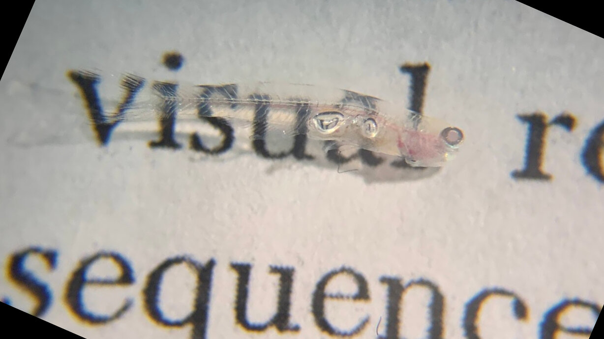
The team profiled more than 600,000 cells taken from the brain, spinal cord, eyes, internal organs, fins, head and trunk. It used single-cell RNA sequencing to measure gene activity in individual cells, then paired those measurements with spatial methods that mapped cell types and gene activity back to their locations in tissue. The fish, which the release says is barely one centimetre long and was formally described in 2021, made the experiment possible because of its size and its close relationship to zebrafish.
What the atlas showed
Across the nervous system, neurons from the same region tended to have more similar patterns of gene activity than neurons that used the same neurotransmitter but sat in different regions, a pattern the release says has also been seen in other vertebrates including mammals. Connective-tissue cells called fibroblasts kept region-specific activity of genes known for establishing different parts of the body during development, and cells in the pectoral and pelvic fins retained gene programs associated with forelimb and hindlimb development. The release states that what these persistent signals do in adult fish is not known.
Injury response and a public resource
After the researchers injured the telencephalon, a region of the forebrain, the missing neurons regenerated, and the team traced a progression from an early wound and immune response through neural stem-cell activity to the production and maturation of new neurons.
The atlas has been made publicly available through DigiFish, an interactive resource for exploring cell types and gene activity across the animal, the institute said.